A new, state-of-the-art microscope will give researchers the tools they need to advance cutting-edge work into cancer treatments.
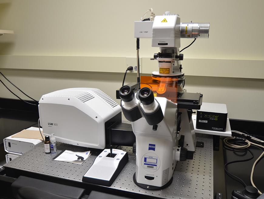
Last week, The Hormel Institute, University of Minnesota, announced it had acquired a $350,000 Zeiss LSM 900 confocal microscope. Confocal microscopes use a system of pinholes to remove out-of-focus light, resulting in sharper, higher-quality images that allow researchers to distinguish between objects that are very close together. The new instrument, which weighs 600 pounds and was shipped from Germany, was funded through support from The Hormel Foundation and the UMN Office of the Vice President for Research.
The LSM 900 provides a major upgrade over the institute’s previous confocal microscope, which was nearing the end of its lifespan and was not functioning properly. Todd Schuster, the institute’s instrument core facility manager, said the new microscope is already being used by many different labs.
“A lot of researchers use this instrument to continue research that they haven’t been able to do with our older system,” he said. “Scientists are using online training to refine their skills and learn newer methods of imaging possible with this microscope.”
The confocal microscope’s ability to fine-tune its focus within a sample helps scientists see different events happening in the same cell. It can also combine images from different levels of the sample to form a 3D image of the specimen being studied.
Sergio Gradilone, PhD, associate professor and head of the Cancer Cell Biology and Translational Research lab, said the microscope will play an integral role in his lab’s work. Gradilone’s team focuses on the biological role of primary cilia—the “antennae” that protrude out from the body of a cell into its surrounding environment—on cancer development. His team found these cilia suppress cancer growth, but many cells at the site of cancer in the body are missing their primary cilia. Understanding how and why ciliary loss happens may help in developing treatments that restore primary cilia as a defense against cancer.
In collaboration with Liqiang Chen, PhD, from the Center for Drug Design in the UMN Twin Cities College of Pharmacy, Gradilone is now working on a pilot project funded through the Masonic Cancer Center’s ChainBreaker grant to develop treatments that simultaneously restore cilia and halt cancer growth. The new microscope will let his team assess different drug candidates and more easily and accurately quantify how cilia are being restored.
“Having technology such as this is extremely important to further successful outcomes for the researchers at The Hormel Institute,” Gradilone said. “The new confocal enables precise, three-dimensional imaging, improved analyses of cellular organelles, and intracellular localization of proteins so we can study more accurately how cancer cells develop and metastasize, leading to more knowledge about how to prevent and control cancer.”